

What Does The Lysosome Do In The Animal Cell
Lysosomes are membrane-enclosed organelles that contain an assortment of enzymes capable of breaking down all types of biological polymers—proteins, nucleic acids, carbohydrates, and lipids. Lysosomes role as the digestive arrangement of the prison cell, serving both to degrade material taken up from exterior the cell and to assimilate obsolete components of the cell itself. In their simplest form, lysosomes are visualized as dense spherical vacuoles, just they can display considerable variation in size and shape as a result of differences in the materials that have been taken up for digestion (Figure ix.34). Lysosomes thus correspond morphologically diverse organelles divers past the common part of degrading intracellular textile.

Figure 9.34
Electron micrograph of lysosomes and mitochondria in a mammalian cell. Lysosomes are indicated by arrows. (Visuals Unlimited/K. G. Murti.)
Lysosomal Acrid Hydrolases
Lysosomes incorporate well-nigh 50 different degradative enzymes that can hydrolyze proteins, DNA, RNA, polysaccharides, and lipids. Mutations in the genes that encode these enzymes are responsible for more than 30 different human genetic diseases, which are called lysosomal storage diseases because undegraded material accumulates within the lysosomes of affected individuals. Most of these diseases result from deficiencies in single lysosomal enzymes. For instance, Gaucher'southward affliction (the most common of these disorders) results from a mutation in the factor that encodes a lysosomal enzyme required for the breakup of glycolipids. An intriguing exception is I-cell illness, which is acquired by a deficiency in the enzyme that catalyzes the first step in the tagging of lysosomal enzymes with mannose-6-phosphate in the Golgi apparatus (come across Figure 9.25). The result is a general failure of lysosomal enzymes to be incorporated into lysosomes.
All of the lysosomal enzymes are acid hydrolases, which are active at the acidic pH (about 5) that is maintained within lysosomes merely not at the neutral pH (about 7.2) characteristic of the rest of the cytoplasm (Figure 9.35). The requirement of these lysosomal hydrolases for acidic pH provides double protection against uncontrolled digestion of the contents of the cytosol; fifty-fifty if the lysosomal membrane were to pause downwardly, the released acrid hydrolases would exist inactive at the neutral pH of the cytosol. To maintain their acidic internal pH, lysosomes must actively concentrate H+ ions (protons). This is achieved past a proton pump in the lysosomal membrane, which actively transports protons into the lysosome from the cytosol. This pumping requires expenditure of energy in the form of ATP hydrolysis, since it maintains approximately a hundredfold higher H+ concentration within the lysosome.
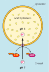
Figure 9.35
Organization of the lysosome. Lysosomes contain a variety of acrid hydrolases that are active at the acidic pH maintained within the lysosome, but not at the neutral pH of the cytosol. The acidic internal pH of lysosomes results from the action of a proton (more...)
Endocytosis and Lysosome Formation
One of the major functions of lysosomes is the digestion of cloth taken up from outside the jail cell by endocytosis, which is discussed in particular in Chapter 12. All the same, the office of lysosomes in the digestion of fabric taken upwards by endocytosis relates not only to the function of lysosomes just also to their formation. In detail, lysosomes are formed by the fusion of send vesicles budded from the trans Golgi network with endosomes, which contain molecules taken upwards by endocytosis at the plasma membrane.
The germination of lysosomes thus represents an intersection betwixt the secretory pathway, through which lysosomal proteins are processed, and the endocytic pathway, through which extracellular molecules are taken up at the cell surface (Figure nine.36). Material from outside the jail cell is taken up in clathrin-coated endocytic vesicles, which bud from the plasma membrane and and then fuse with early endosomes. Membrane components are then recycled to the plasma membrane (discussed in detail in Affiliate 12) and the early endosomes gradually mature into late endosomes, which are the precursors to lysosomes. One of the important changes during endosome maturation is the lowering of the internal pH to well-nigh 5.5, which plays a key role in the delivery of lysosomal acid hydrolases from the trans Golgi network.
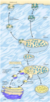
Figure 9.36
Endocytosis and lysosome formation. Molecules are taken upward from exterior the cell in endocytic vesicles, which fuse with early endosomes. Membrane components are recycled every bit the early endosomes mature into belatedly endosomes. Transport vesicles carrying acid (more...)
As discussed before, acrid hydrolases are targeted to lysosomes past mannose-6-phosphate residues, which are recognized by mannose-vi-phosphate receptors in the trans Golgi network and packaged into clathrin-coated vesicles. Post-obit removal of the clathrin coat, these transport vesicles fuse with late endosomes, and the acidic internal pH causes the hydrolases to dissociate from the mannose-6-phosphate receptor (see Figure nine.36). The hydrolases are thus released into the lumen of the endosome, while the receptors remain in the membrane and are eventually recycled to the Golgi. Tardily endosomes then mature into lysosomes as they larn a total complement of acrid hydrolases, which digest the molecules originally taken upward by endocytosis.
Phagocytosis and Autophagy
In add-on to degrading molecules taken up by endocytosis, lysosomes digest fabric derived from two other routes: phagocytosis and autophagy (Figure 9.37). In phagocytosis, specialized cells, such as macrophages, take upwardly and degrade large particles, including leaner, jail cell debris, and aged cells that need to exist eliminated from the torso. Such large particles are taken up in phagocytic vacuoles (phagosomes), which then fuse with lysosomes, resulting in digestion of their contents. The lysosomes formed in this way (phagolysosomes) can be quite large and heterogeneous, since their size and shape is determined past the content of material that is beingness digested.
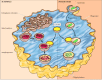
Effigy 9.37
Lysosomes in phagocytosis and autophagy. In phagocytosis, large particles (such as bacteria) are taken up into phagocytic vacuoles or phagosomes. In autophagy, internal organelles (such every bit mitochondria) are enclosed by membrane fragments from the ER, (more...)
Lysosomes are also responsible for autophagy, the gradual turnover of the cell'southward own components. The first step of autophagy appears to be the enclosure of an organelle (e.g., a mitochondrion) in membrane derived from the ER. The resulting vesicle (an autophagosome) then fuses with a lysosome, and its contents are digested (see Effigy ix.37). As discussed in Chapter vii, autophagy is responsible for the gradual turnover of cytoplasmic organelles.
![]()
Box
Molecular Medicine: Gaucher's Disease.

Source: https://www.ncbi.nlm.nih.gov/books/NBK9953/
Posted by: youngwermell.blogspot.com
0 Response to "What Does The Lysosome Do In The Animal Cell"
Post a Comment